Optical tomography is a novel method for biomedical imaging applications which is rapidly gaining importance. This technology uses near-infrared (NIR) light from a single or several sources to illuminate a specimen. Multiply-scattered light which emerges at a different location is observed using a detector unit. A model of the light propagation physics (the so called forward model) is utilised to solve the inverse imaging problem. This means the localized optical properties (absorption or scattering coefficients) of the investigated tissue will be reconstructed and can be shown in the form of a cross sectional view. Currently the main biomedical applications for optical tomography are tumor detection in the female breast and imaging of the brain.
In the presented work an optical tomography system, which has been developed during a doctoral thesis, is in-depth analysed with measurements. The tomography system is based on a frequency-domain method; this means intensity modulated light is coupled into the surface of the medium under investigation. The detected signal, which is collected at a different surface location, is of the same frequency but amplitude damped and phase shifted. These amplitude ratios and phase differences, measured at several surface positions, contain information about the optical properties of the medium. Inhomogeneities that have absorption or scattering contrast to their surrounding medium can be detected. A characteristic of the measurement system is that it has just one source and one detector fibre which are integrated in a measurement-head and scan two dimensionally with a traction unit over the surface of the object.
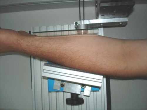
Measurements with specially produced technical (resin) phantoms and in-vivo analyses have been accomplished. The in-vivo measurements were focused on the detection of a known lipom in the forearm of a test person. Based on new insights some optimisations were put in place that simplify the operation of the tomography system and assure a high quality of the results. These include the programming of graphical user interfaces (GUI) for data aquisition and reconstruction and finding appropriate parameter sets to obtain optimal measurement and image reconstruction results.
February 2006
 Optical tomography is a novel method for biomedical imaging applications which is rapidly gaining importance. This technology uses near-infrared (NIR) light from a single or several sources to illuminate a specimen. Multiply-scattered light which emerges at a different location is observed using a detector unit. A model of the light propagation physics (the so called forward model) is utilised to solve the inverse imaging problem. This means the localized optical properties (absorption or scattering coefficients) of the investigated tissue will be reconstructed and can be shown in the form of a cross sectional view. Currently the main biomedical applications for optical tomography are tumor detection in the female breast and imaging of the brain.
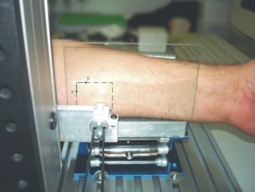
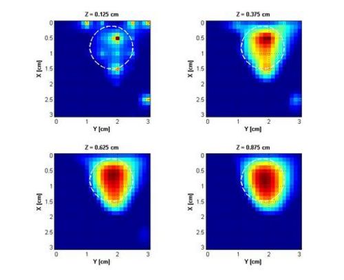 Abbildung 2: Rekonstruierte Schnittbilder einer Lipom–Detektions–Messung; die tatsächliche Lage bzw. die ungefähre maximale Ausdehnung des Lipoms sind strichliert eingezeichnet.
Johannes Kepler University Linz
Altenberger Straße 69
4040 Linz, Austria
 Go to JKU Homepage
