A major topic in the investigation of nanostructures with x-rays is the use of local resolution. Usually, the x-ray beam diameter is much larger than the structures, and hence a large ensemble, often consisting of millions of individual nanostructures, is illuminated. This is all right to obtain the structural properties with a high statistic relevance, but the approach fails for inhomogeneous samples with a large spread of properties, where no meaningful "average structure" exists. This is the case for samples with several different kinds of nanostructures, or processed samples, where one wants to investigate only a certain part of the sample.
A way to address this problem is to use focused x-ray beams. Since most materials have a refractive index very close to unity for x-rays, this cannot be done by classical "lenses". Several concepts can, however, be used to focus x-rays. We are mainly using three of them, in collaborations with synchrotron sources:
Fresnel zone plates, with the zones alternatingly transparent or opaque for x-rays can be used the same way as in conventional optics. To achieve focusing in the 100 nm range, outer zone diameters must be in the same range, putting a limit to the thickness of material used (often gold). Hence the "opaque" zones are in fact not opaque, but fabricated in a way to cause a phase shift of pi for transmitted x-rays.
Mirrors can also be used for x-rays, but the incidence angles have to be very glancing with respect to the surface. They have to be very smooth and are often fabricated from silicon, eventually coated with heavier elements. To ensure the correct parabolic profile, often two mirrors shaped like a parabolic prism are used in a crossed arrangement (to be able to focus horizontally and vertically), also called "Krickpatrick-Baez" setup.
Actually, refractive lenses of materials with low absorption (usually Be for x-ray energies below 12 keV, or Si for higher energies) can be used. Sinca a single lens does not defract enough, multiple (about 10 to 100) lenses are stacked into so-called "compound refractive lenses" (CRL). The focal length can be tuned by the number of single lenses.
With the concepts above, focal lengths in the order of few 10 cm can be achieved. To obtain a really small focus, already small source sizes have to be demagnified, so large distances to the source are required (50-100 m typically). This is only possible at synchrotron sources, which in addition offer the required photon flux and brilliance for experiments with focused beams.
performing the measurement
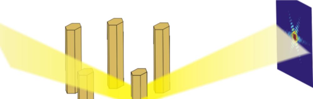
Producing a focused x-ray is one thing, illuminating a specific single nanostructure on a sample, another one. The first problem one encounters is to know where the focused beam hits the sample. Using standard alignment procedures of the focus to the center of rotation of the goniometer, and of the sample into this center of rotation, usually is limited to a precision of few micrometer.
For more precise alignments, the sample has to be aligned using the focused x-ray beam, i.e., a diffraction signal typical for the nanostructure(s), which are to be investigated, has to be found. Then the intensity can be recorded as a function of lateral sample position, to map out the real-space distribution of nanostructures, and to identify the particular one for a detailed analysis.
Unfortunately, this is not the end of the story: in a diffraction experiment, the sample is typically rotated by few 0.1 degrees around a Bragg peak position, and measurements around several Bragg peaks, often apart by few 10 degrees, need to be recorded. Since no bearings for rotation axes with a precision in the 10 nm range, with the required large travel ranges exist, this means that the alignment has to be repeated for every Bragg peak. Only at very dedicated instruments the precision of the goniometer is sufficient to at least keep the focused beam in the same spot (within few 10 nm) during the scan at a single Bragg peak.
A further annoyance are vibrations, omnipresent at synchrotrons with a lot of machinery around a beamline, and temperature drifts. To keep a position within few 10 nm for hours of an experiment, special care has to be taken to eliminate vibrations (mostly by using large masses as base of a goniometer, decoupling it from the ground, but rigidly coupling it to the focusing element) and keep the temperature stable to about 0.1 degrees C.
our project
We have a collaboration with beamline ID01 at the European Synchrotron Radiation Facility (ESRF) in Grenoble, France, establishing a focusing setup for x-ray beam sizes down to 100 nm, at beam energies in the 10 keV range. Currently, we are reaching focus sizes around 300 nm using Fresnel zone plates or CRLs. The focused beams have been used to investigate single epitaxial SiGe islands on Si(001), sinlge InAs and InAs/InAsP nanowires on Si and InP substrates, and also a single SiGe island embedded into a functioning transistor device.
Johannes Kepler University Linz
Altenberger Straße 69
4040 Linz, Austria
 Go to JKU Homepage
