| Name |
Purpose |
Lifetime |
Provider |
|
_gcl_au
|
This cookie is used by Google Analytics to understand user interaction with the website.
|
3
months
|
Google
|
|
_ga
|
This cookie is installed by Google Analytics. The cookie is used to calculate visitor, session, campaign data and keep track of site usage for the site's analytics report. The cookies store information anonymously and assign a randomly generated number to identify unique visitors.
|
2
years
|
Google
|
|
_gid
|
This cookie is installed by Google Analytics. The cookie is used to store information of how visitors use a website and helps in creating an analytics report of how the website is doing. The data collected including the number visitors, the source where they have come from, and the pages visited in an anonymous form.
|
1
day
|
Google
|
|
_gac_UA-112203476-1
|
Contains campaign related information for the user and measures the AdWords campaign success.
|
90
days
|
Google
|
|
test_cookie
|
This cookie is set to determine if the website visitor's browser supports cookies. Doesn't contain personal identifier.
|
15
minutes
|
Google
|
|
IDE
|
This cookie carries out information about how the end user uses the website and any advertising that the end user may have seen before visiting the said website.
|
1
year
|
Google
|
|
_gcl_aw
|
This cookie is set when a user clicks an ad to reach our website. It informs about the success of campaigns and allows to connect ads to conversion targets.
|
3
months
|
Google
|
|
AMCV_xx
|
This is a pattern type cookie name associated with Adobe Marketing Cloud. It stores a unique visitor identifier, and uses an organisation identifier to allow a company to track users across their domains and services.
|
3
years
|
LinkedIn
|
|
bcookie
|
Contains a browser ID.
|
2
years
|
LinkedIn
|
|
bscookie
|
Contains a browser ID for a secure connection.
|
2
years
|
LinkedIn
|
|
lang
|
This cookie is used to store the language preference of our visitors
|
Session
|
LinkedIn
|
|
lidc
|
This cookie carries out information about how the end user uses the website and any advertising that the end user may have seen before visiting the said website.
|
1
day
|
LinkedIn
|
|
lissc
|
This cookie is used to analyze how a visitor interacts with embedded services.
|
1
year
|
LinkedIn
|
|
UserMatchHistory
|
This cookie is set when a user clicks an ad to reach our website. It informs about the success of campaigns and allows to connect ads to conversion targets.
|
30
days
|
LinkedIn
|
|
fr
|
This cookie is set when a user clicks an ad to reach our website. It informs about the success of campaigns and allows to connect ads to conversion targets.
|
90
days
|
Facebook
|
|
fbp
|
This cookie is used to display advertisings, for example third-party real time offers.
|
90
days
|
Facebook
|
|
sc_at
|
This cookie is used to identify a visitor across multiple domains.
|
1
year
|
Snap
|
|
sc-country
|
This cookie is used to determine a visitor's country.
|
1
day
|
Snap
|
|
uid
|
This cookie sets a random User-ID and helps at real time bidding for display advertising to targeted audiences.
|
60
days
|
Adform
|
|
C
|
This cookie identifies if user’s browser accepts cookies. 1 – Cookies are allowed, 3 – Opt-out.
|
30
days
|
Adform
|
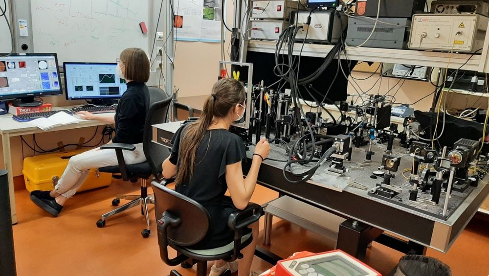 AO-OCT setup used to perform retinal imaging on a healthy volunteer.
©Medical University of Vienna
AO-OCT setup used to perform retinal imaging on a healthy volunteer.
©Medical University of Vienna
 Go to JKU Homepage
