Go to JKU Homepage

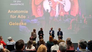
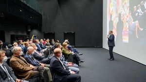
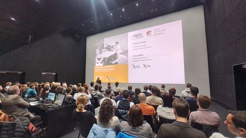
The presentation "Radiology and Anatomy - Past, Present, and the Future" at the JKU's medSPACE took the audience on a journey through the history of radiology.
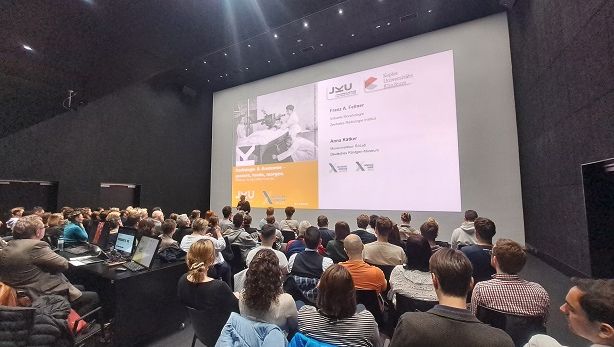
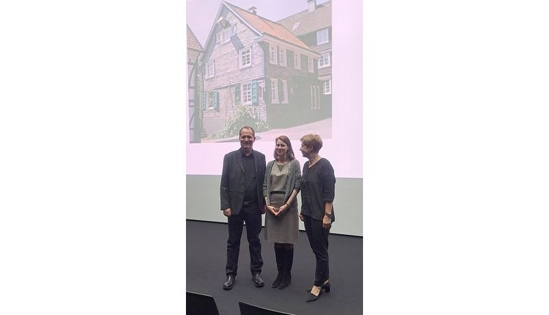
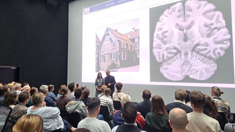
Guest speaker Anna-Katharina Kätker (of the German Röntgen Museum located in Wilhelm Conrad Röntgen's hometown of Remscheid) took the audience on a journey through time, starting at the discovery of X-rays by the physicist Röntgen, to subsequent advancements resulting from its initial popularly and then, oftentimes, unfortunate ramifications of the new technology. It is a story shaped primarily by women at the forefront who enthusiastically pioneered radiology technology and whose importance was seldom appreciated in the past. Kätker provided an overview of the subject area’s educational background up to creating the Deutsches Röntgen-Museum Remscheid and the educational concept behind the museum.
Prof. Franz Fellner continued with a presentation about the current technologies of computer tomography (CT) and magnetic resonance imaging (MRI), both of which are diagnostic imaging procedures.
One of the JKU's key strengths is creating and expanding on the award-winning "cinematic rendering" process, a three-dimensional representation of a living human body and its unique educational applications to teach the course "Virtual Anatomy and Patho-Anatomy" at the JKU's Faculty of Medicine, thereby replacing the otherwise standard cadaver-based approach to teaching.
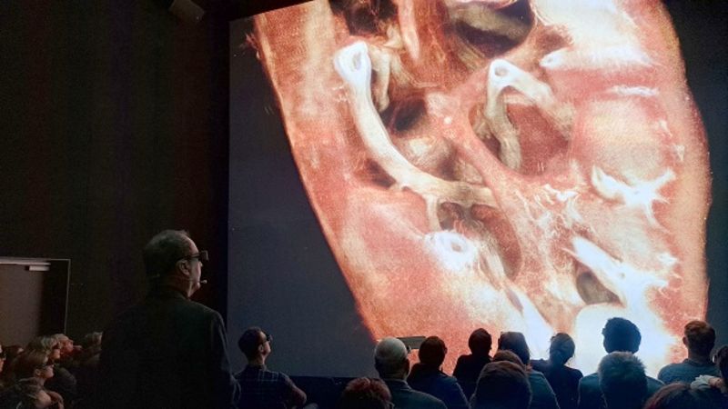
The audience members also shared a glimpse into the future along with Prof. Fellner as he showcased the latest datasets of kidneys, brains and hearts (donated organs) created using the synchrotron (particle accelerator) in Grenoble. A presentation highlight included a 3D "mesoscopic" (between macroscopy and microscopy) view of a heart and its ventricle in combination (and in 3D) with a video recording of an operation performed by Univ. Prof. Dr. Andreas Zierer and focusing on this exact area of the heart valve and the ventricle during the operation.


![[Translate to Englisch:] Eindrücke von Anatomie für Alle](/fileadmin/_processed_/3/8/csm_JKUmedSPACE_VirtuelleAnatomie_02_2000px_72e7ba2fbc.png)