 Go to JKU Homepage

The HistoCore Arcadia H is a modern paraffin embedding station from Leica. It is used for embedding histological tissue samples, after fixation, into liquid paraffin. Before solidification into paraffin the tissue is properly oriented with tweezers.
The Core Facility Histology operates all necessary equipment for the traditional preparation of histological samples at the Center for Medical Research, for processing at room temperature (FFPE samples) as well as at low temperatures (cryofixation). It is equipped with modern imaging techniques and scanners that enable rapid analysis in brightfield and fluorescence, as well as the identification of over 100 biological markers in a single tissue section. In addition to training on equipment, the Core Facility Histology also offers consultations in the areas of sample preparation, sample processing, and image analysis.
The HistoCore Arcadia H is a modern paraffin embedding station from Leica. It is used for embedding histological tissue samples, after fixation, into liquid paraffin. Before solidification into paraffin the tissue is properly oriented with tweezers.
The HistoCore Arcadia C is a cooling plate from Leica that can be used for both cooling and curing of paraffin tissue blocks.
The RM2245 from Leica is a semi-automatic rotary microtome used for cutting formalin-fixed, paraffin-embedded histological tissues, particularly for users who prefer manual cutting.
The HistoCore AUTOCUT is a motorized rotary microtome that can be used both manually and fully motorized. This device is capable of cutting formalin-fixed paraffin-embedded tissues of various hardness grades at different section thicknesses.
The Leica CM3050 S is a semi-motorized high-performance cryostat with the option for motorized cutting. It is used to rapidly freeze fresh histological samples and cut them while in a frozen state.
The Leica VT1200 S is primarily used to cut fresh biological tissue in a cooled buffer. The goal is to preserve the integrity of the tissue cells for further biological processes. Cutting with the VT1200 S is motorized.
A multifunctional microwave tissue processor for the processing and fixation of histological tissues. Additional procedures such as antigen retrieval and decalcification of bones can also be performed.
Microwave Tissue Processor KOS, opens a file in a new window
The Olympus VS200 Slide Scanner supports brightfield and fluorescence scans (DAPI, CY3, CY5, CY7, and FITC) on up to 6 different histological samples simultaneously.
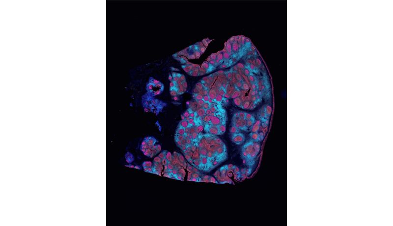
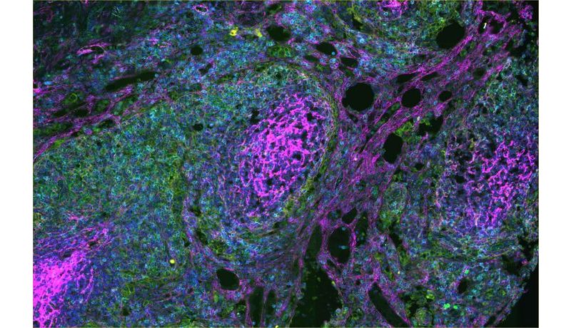
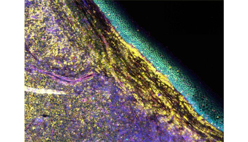
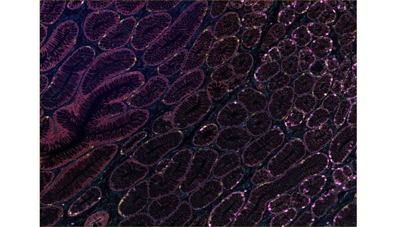
The PhenoCycler-Fusion System by Akoya BioSciences enables spatial ultra-high-plex phenotyping of entire slides with single-cell resolution through automated fluidics and iterative imaging. The automated cycling and image capture are facilitated by the integration of flow cells placed in a slide holder. This allows the detection of over 100 markers on a single intact tissue. This device is operated by the Core Facility and is not independently operated by users.
PhenoCycler-Fusion System Akoya, opens a file in a new window

Name | Position | Phone | E-mail Address |
| Claudia Himmelbauer, MSc | Head of Core Facility | 0732 2468 3093 0664 602468213 | claudia.himmelbauer@jku.at |
Johannes Kepler University Linz
Altenberger Straße 69
4040 Linz, Austria